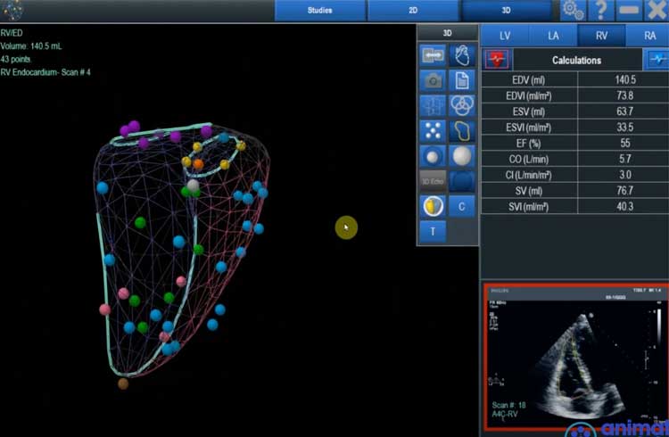
With our patented A.I., ultrasound scans are turned into high-fidelity 3D models of all four chambers of the heart, at a fraction of the cost and time of a traditional MRI.
Ventripoint is now being used by major hospitals in Europe, the U.K., the U.S. and Canada.
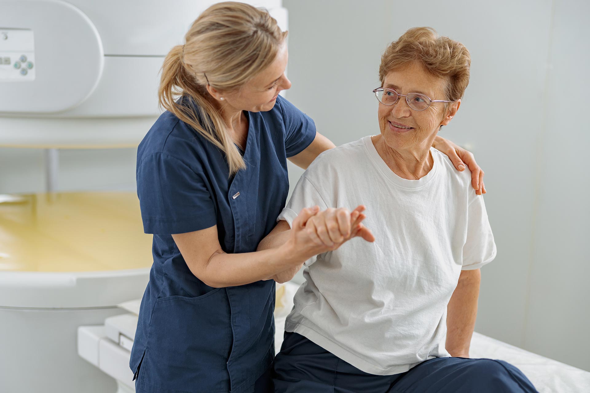
Our VMS+ is a diagnostic aid that was developed to provide a point of care solution to better communicate the heart’s structure and function without the need for MRI.
The VMS+ marries the ingenuity of the cardiac MRI and Echo with the power of Ventripoint’s Knowledge Base Reconstruction (KBR). KBR powers construction of a 3D model of the heart and calculates volumes and ejection fractions for all chambers with an accuracy comparable to the MRI.
The British Heart Foundation & iTV Global Report on Ventripoint
Products
Our Breakthrough Technology
VMS+
Harness the Power of KBR
VMS+ is designed to create an accurate 3D model of the heart from a 2D echocardiogram to obtain cardiac metrics on all four chambers of the heart within minutes. Obtain accurate results that do not require “perfect images”.
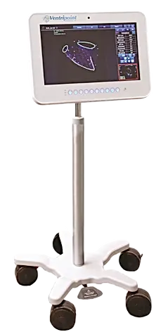
VMS+ software can be installed on any personal workstation. Create a 3D model of the heart’s chambers with the use of any commercially available 3D echo or MRI images. Obtain reproducible and accurate volumetric measurements and ejection fractions.
"Having had experience with the older Ventripoint system, the new VMS+ system with its increased functionality is a great addition to our echo lab.”
“We want to introduce Ventripoint to all our hospital partners, to make it a standard of care,”
"VMS+ not only gives us a very precise spatial view of complex congenital malformations at any time, but it also provides us with important data on the function of the heart muscle. This allows us to plan therapy even more precisely, check it at any time and adjust it if necessary,”
“Our aim is that by working together, we can ultimately provide reliable and effective tools for clinicians on a global scale.”
"Having had experience with the older Ventripoint system, the new VMS+ system with its increased functionality is a great addition to our echo lab.”
“We want to introduce Ventripoint to all our hospital partners, to make it a standard of care,”
Want to talk to us about VMS+?
Talk to a representative to learn how VMS+ can be used to aid in more accurate volumes and ejection fractions so you can follow patients with confidence.