

Dr. George Adams
January 13, 2022
The Future is Fast Approaching
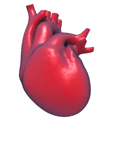
Ventripoint’s development roadmap is firmly set on achieving the goal of a fully-automated analysis of the whole heart from all 2D and most 3D echocardiograms. We already have shown we can semi-automatically analyze even the poorest 2D scans and fair 3D scans for all 4 chambers of the heart. We have developed working prototypes to do this automatically for 2D and 3D datasets. We anticipate we will have the final product for 2D and 3D scans this year.
The dream in heart analysis has been to reduce the time to “read the heart” from 30-45 minutes by automatically analyzing a single holographic picture of the heart (3D ultrasound). 3D ultrasound was a big hit with the “baby face” application where you could get a look at an unborn fetus in the womb and see the child’s face in 3D. Obtaining a good picture of the heart has been getting better over the last 15 years, but it is still not “good enough” to be analyzed routinely using conventional techniques. The images are still blurry and incomplete and so too often unreadable. Still the dream continues.
With the backlog of heart patients from the curtailment of echocardiography due to the COVID pandemic (estimated to be 6-36 months to clear) and the new long-COVID patients with cardiac dysfunction, the need has never been higher. We are on the cusp of achieving the dream.
There will be 25% of patients who will still need 2D exams as 3D echocardiography will still not work for these people. Hence, we and a panel of leading cardiologists have imagined an even better future – one where the automated analysis includes not only volumes and ejection fractions, but motion – shape, wall motion, strain, twist, rates, etc. for chambers as well as for portions of the chambers. This kind of detailed analytics can be done easily once the full dynamic mathematical model of the whole heart is obtained. Once again, we have these all ready to be implemented in the next generation. This is new version will also include an upgraded tracking system, which is compatible with the pacemakers and defibrillators, which are often implanted into heart patients. Soon everyone’s heart can be scanned and read.
After decades of ultrasound manufacturers improving 3D ultrasound machines and Ventripoint improving the analytics, it is now time to put it all together. It will not be long. The future is approaching fast!
Dr. George Adams
October 22, 2021
The Way We Measure High Blood Pressure Makes My Blood Boil
.png)
The WHO just released a comprehensive set of guidelines for treating hypertension and reports “An estimated 1.4 billion people worldwide have high blood pressure, but just 14% have it under control. However, cost-effective treatment options do exist.”(1) The WHO’s definition of hypertension is ≥140/90 mmHg which is higher than the current guideline from the American Heart Association which is ≥130/80 mmHg (2). By this definition, 46% of Americans have hypertension. (3) People with this level of high blood pressure have double the risk of cardiovascular complications compared to those with a normal level of blood pressure (120/80 mmHg). In Canada, a recent commentary by the experts in hypertension stated, “After more than 60 years of declining rates of cardiovascular death, trends are now reversing, along with reduced rates of detection, treatment and control of hypertension”. (4)
Many studies have shown even a small decrease in hypertension has great benefits to the patient and reduce the cost of cardiovascular disease to a healthcare system. (1) Best practices call for a “treat-to-target” approach, meaning set a target for each patient and treat them aggressively through life-style changes and drugs to get to that target.(1) An interesting study just published in the NEJM found more aggressive treatment with a target of 110 mmHg in elderly patients with hypertension resulted in many fewer cardiovascular events, including death over a 4-year period than standard target of <130mmHg. (5) The lower the better and we have cheap ways, like life-style changes and medications to lower blood pressure.
All the evidence for decades has shown lowering blood pressure is beneficial so why is it not practiced? It is complicated but identifying people with chronic hypertension is certainly the first step that needs to be improved. Most doctors use the blood pressure cuff technique and while the guidelines call for the patient to sit quietly in a chair for at least 5 minutes prior to the test, this is seldom done in a busy GP’s office. The result is doctors often see high blood pressure measurements and just discount them as “white-coat” hypertension – high due to being at the doctor’s office.
Hypertension is the most common reason to visit a doctor; in 2007, 21.1 million visits to community physicians in Canada were made for high blood pressure. (6) Everyone’s blood should boil at the inefficient way we diagnose and NOT treat hypertension, so we can pay 100 times more on emergency care. If ever there was a place where an ounce of prevention saved a pound of cure, this is it.
1. https://apps.who.int/iris/bitstrea m/handle/10665/344424/9789240033986-eng.pdf
2. https://www.heart.org/en/health-topics/high-blood-pressure/the-facts-about-high-blood-pressure
4. https://hypertension.ca/wp-content/uploads/2021/06/Leung-refocusing-htn-ctrl-CMAJ2021.pdf
Dr. George Adams
September 13, 2021
Remembering the Forgotten Ventricle in Unforgettable People

It has been eleven years since Dr. Luc Mertens from the Hospital for Sick Children wrote a review in Nature Medical Journal* stating, “The right ventricle of the heart has long been the ‘forgotten ventricle’, as it is difficult to image…” For people born with a heart problem, a condition known as Congenital Heart Defect or CHD, it is a lifelong issue. From the very beginning of life for people with CHD, cardiologist need to be able to view and assess the Right Ventricle (RV) frequently to plan the next surgery or a change in treatment at the optimal time. The RV is renowned for its inability to recover once it has been stretched too far for too long and so finding the last responsible point to treat CHD patients is an all-encompassing effort for clinicians. The difficulties of imaging the RV results in traditional ultrasound being inadequate in 20-50% of CHD patients and so expensive and complex MRI is required.
This has all changed with Ventripoint’s novel Artificial Intelligence (AI) approach to analyze the RV. The Company’s VMS+ 3.0 analytical software offers a proven, patented, non-invasive AI solution for 3D visualization of all four chambers of the heart, providing a pivotal new tool for cardiologists to combat cardiovascular disease and sustain heart health in both adults and children. VMS+ 3.0 delivers a more precise and economical way to image the heart, providing results equivalent to more costly and time-consuming MRI. These advantages also allow for faster and more frequent exams.
VMS (Ventripoint Medical System) marries the ingenuity of cardiac MRI (Magnetic Resonance Imaging) through reference databases and ultrasound with the power of Ventripoint's proprietary KBR (Knowledge-Based Reconstruction) AI technology. KBR enables physicians to construct a precise 3D model of the heart and calculate volumes and ejection fractions for all chamber of the heart with an accuracy and reliability equivalent to MRI.
The Company now has a dozen studies underway or in the planning stage in top cardiac centres to show the RV is important in all types of heart disease (not just CHD). A particularly unique study is assessing mothers’ hearts during pregnancy, when there is a 50% increase in load on the heart**. Women with known heart conditions, like CHD, are considered high-risk and are seen frequently in the last trimester of the pregnancy as once again it is the RV that is most stressed and likely to fail. After all, moms are the most unforgettable people we know, and now cardiologists have a way to image these life-producing precious hearts.
* Mertens LL, Friedberg MK. Imaging the right ventricle--current state of the art. Nat Rev Cardiol. 2010 Oct;7(10):551-63. doi: 10.1038/nrcardio.2010.118. Epub 2010 Aug 10. PMID: 20697412.
**Ramlakhan, K.P., Johnson, M.R. & Roos-Hesselink, J.W. Pregnancy and cardiovascular disease. Nat Rev Cardiol 17, 718–731 (2020). https://doi.org/10.1038/s41569-020-0390-z
Dr. George Adams
February 8, 2021

It is amazing to witness the evolution of a product like the VMS+ from its beginning stages to the latest version we have today. Like any product development, growth comes from seeing what can be improved, removed, added, and adjusted. But most importantly, growth comes from experience - our user’s experience.
In order to put out the best product possible, we like to check in with our customers on how their experience with using the VMS is going, and what changes can be made to improve user experience and efficiency. The feedback we receive from our users is critical to us, because it’s how we shape and improve the VMS+.
The evolution of the VMS+ over the last decade is apparent when looking at the first VMS system. But aside from the system's appearance, how we fit into a clinician’s appointment evolved over time. A key piece of feedback we received that we used during development was that the VMS needs to fit into the standard workflow. Since the clinician has only about 45 minutes to complete a full echo exam, our system couldn’t take up any extra time. We designed the system to be part of the standard exam which required us to only use standard images that conform to DICOM standards. With the information we have from our user’s experience, we can keep improving the VMS+ and make it a valuable asset for assessment.
The VMS+ will continue to develop with the feedback we receive from our users today. The ability to make appointments easier for both clinicians and patients is what we strive for and the reason why feedback is so important to us.
Dr. George Adams
November 24, 2020
Why are we doing this?
My job is a mix of talking to potential investors, existing shareholders, stock analysts and potential partners, as well as encouraging everyone within the Company to focus on execution. For me, it is all the same message: “why are we doing this?”. It is all about making a difference – making the world a better place. I have very little time for shareholders who only want to make a small return on their investment by timing the market. Don’t get me wrong, I am all for making a large return. This is my 7th company as CEO and they have all made money for investors who stayed the course. It is about building a real company with a real solution to a real problem. Yes, it has taken a number of teams of people over 10 years to perfect the VMS technology, but we have it now in the VMS+3.0, and just in time, as COVID-19 patients are experiencing heart damage on an unprecedented scale. 60% have acute cardiac dysfunction and many of those go on to have continuing problems – “the long haulers” as they are called. There are social networks groups with thousands of members sharing their chronic symptoms post-COVID and looking for answers. It feels good to have a diagnostic tool to offer them.
Often investors ask me “why I am doing this?” I have participated in the development of products that have extended the lives (nobody ever saves a life) of 300 million people in my 35 years as a serial entrepreneur. Sometimes, I think “I have done my bit”, but then I realize there is more to be done. Heart disease is still the number one health problem worldwide. I remember attending the World Economic Forum many years ago and listening to a presentation describing the rapid rise of cardiovascular disease in the developing countries of the world, who had managed to decrease deaths from infectious diseases. This advancement allowed their citizens to live longer and unfortunately develop heart disease.
I do this because I can – I can build a team to make a difference worldwide by developing a technology that works with 2D ultrasound, which is used worldwide already, to allow the correct diagnosis and monitoring of people with heart disease. I can inspire investors and shareholders to see the big picture and be patient. That is my superpower! It helps they have seen this movie 6 times already and even though Ventripoint has taken over a decade to mature, I am pleased to say, I have lots of shareholders who have been with me from the beginning. The Company has never been stronger. It has never been more able to address the needs of children and adults with heart disease.
That is why we do this.
Dr. George Adams
Oct 6, 2020
The report from Israel of the dilation of the right ventricle (RV) in 39% of patients with COVID-19 is very troubling to cardiologists, who understand the capabilities and limitations of the RV. Both the left ventricle (LV) and RV naturally dilate in response to increased load (pressure or blood volume) or muscle damage due to an infection (virial or bacterial) or a heart attack (vasospasm, myocardial infarction, etc.). When the initial cause is alleviated either naturally or by therapy, the LV can shrink back to a normal size, a process called “remodeling”, and continue to function normally until the next event. However, the RV is not so elastic and once it has been dilated too much for a period of time, it cannot remodel. The literature has documented this phenomenon in patients with congenital heart disease, whose pulmonary valves often fail in their teenage years and as the valve fails, the RV dilates due to the backflow of blood (regurgitation) into the RV. Cardiologists have learned to replace these valves before the RV gets too large.
Patients with pulmonary hypertension have the same fate due to the increased pressure from increased resistance to blood through the lungs for a number of underlying causes. So, when it was observed a large fraction of people infected with COVID-19 had at least an acute dilated RV, it set off the alarm bells for cardiologists. What is needed now is detailed follow-up studies to monitor patients and accurately calculate the volume of the RV over time to see if this effect will be reversed or be permanent. The concern is that this dilation in COVID patients will persist and there will be a tsunami of patients with right heart failure in the coming years.
The VMS+3.0 is the only tool available to accurately and rapidly determinate the size of the RV from a 2D echocardiogram. It would seem our device will be needed more than ever as the pandemic spreads
How big is your heart?
Dr. George Adams
Sept 23, 2020

COVID carnage of the heart was the headline.
ET TU, COVID?
As a scientist, I spent many years studying cells and their cytoskeletons to try to understand how they moved. I made time-lapse videos of them - showing they are always moving - looking to fill in where they are needed. Like a human body, a cell without cytoskeleton and muscle cannot do much but be a blob. So when I saw the amazing pictures of heart cells that had been infected with COVID and the destruction of their cytoskeletons and muscle fibers, I was very concerned what this could mean. Sure it was just an experiment in a petri dish, but the article also found similar disrupted heart cells in post-mortem samples from COVID patients. So it is not so surprising that 50% of people with COVID have heart disfunction, even if they had mild to no symptoms. Never before has such a carnage been observed. This COVID virus is special in its ability to attack the heart's muscle.
We are now hearing of 35% of COVID patents reporting continuing and chronic problems associated with cardiovascular complications. So as you would expect some people overcome the heart damage while others struggle. I can see now why cardiologists are concerned about the possibility of patients with heart problems that go on for decades.
I hope we can assist in some way with the VMS+ 3.0’s unique ability to quickly, accurately and reproducibly analyze the heart and allow for the chronicling of the progression of the heart in healing or continued decline. It seems we are at the right place at the right time to help.
Dr. George Adams
September 10, 2020
PAST, PRESENT, FUTURE

I was attracted to the Company in 2010 as the first VMS1.0 was being clinically evaluated. Dr. Mertens, who is one of the top paediatric cardiologists in the world, had just written a review on the “forgotten ventricle” and published it in Nature*. Everyone was beginning to understand that only analyzing the left side of the heart was ignoring half of the information about how the heart was functioning. 10 years ago only MRI could give you right-heart information and as a cardiologist told me at that time, it was traumatic for the doctor, the parents and the child to suggest a child or infant have an MRI. From then on, I was dedicated to creating the VMS to address this need.
I am often asked if 10 years later, I regret this decision and I certainly do not. Back in the early 80s I invented an artificial medium for platelet storage and licensed it to a major blood transfusion company. It is just now becoming the standard, 35 years later! Significant change in medical practice takes a long time. The promise of 3D ultrasound being able to routinely yield useful information for the right heart has not materialized and so the VMS approach remains the only way to get reliable accurate measurements without waiting 3 months for an MRI appointment and spending at least an hour in the machine.
Ventripoint really pioneered the use of artificial intelligence (AI) with its KBR approach and no-one has been able to find another way to get MRI-grade measurements from regular 2D ultrasound exams. Yes, it has taken a lot of time and money to perfect the VMS+3.0 so it is user-friendly and fits exactly into the workflow of an echocardiography service, but now we have it and are set to change the world and stop the trauma.
*Mertens, L., Friedberg, M. Imaging the right ventricle—current state of the art. Nat Rev Cardiol 7, 551–563 (2010). https://doi.org/10.1038/nrcardio.2010.118
Dr. George Adams
August 14, 2020

BATTER UP
COVID?
A major study published in one of the top medical journals (JAMA) reported COVID damages the heart even in asymptomatic patients. No one really knows how long lasting this damage will be, but Boston Red Sox left-hander Eduardo Rodriguez will miss the entire season because of heart inflammation caused by COVID-19.
Red Sox chief baseball officer Chaim Bloom said "We were optimistic that it would resolve in short order and that we would be progressing back to pitching. As we've continued to monitor it, it has not resolved. It is still there.”
The cardiology community is now very concerned that there will be a tsunami of heart patients due to COVID on top of the regular heart patients who have been waiting months to have their regular echo done. Consequently, they are looking for new faster and better ways to monitor patients. Obviously, the Ventripoint VMS+3.0 can help in this situation as few people have immediate and repeat access to an MRI to document heart damage. We are also hearing from cardiologist about a concern about the backlog and the future where it will be near impossible to have a MRI done as the machines will be needed to deal with other patients. With echo departments near capacity prior to COVID shutdowns, the future looks ominous.
Ventripoint has 4 leading cardiac teams around the world using our latest prototype VMS system to study COVID patients and determine the best way to monitor their progress. It is early days, but the more accurate and reproducible analysis the VMS provides will be needed to identify positive or negative changes in heart function as quickly as possible.
Ventripoint is in this with all of you!
Be healthy and safe!
